Photoclinic
A 16-month-old girl was initially brought to her primary care physician because of persistent nonproductive cough of 1 to 2 weeks’ duration, with lethargy, poor feeding, and worsening cough for the past 36 hours. She had been afebrile. The patient was noted to be pale and had a decreased level of interaction. She was promptly sent to the local emergency department. Laboratory studies showed a hemoglobin level of 3.8 g/dL and hypochromic microcytic anemia. The patient was subsequently transferred to the pediatric ICU for evaluation.
On admission, the infant was pale and tachypneic. Four liters of oxygen were given via nasal cannula. After further questioning of the parents, it was learned that the infant was fed solely cow’s milk and tolerated it well. The primary care physician had had no concerns about weight loss or failure to thrive. There was no history of recurrent otitis media or chest infections.
Chest examination revealed a quiet precordium with normal breath sounds bilaterally. A chest radiograph showed bilateral pulmonary infiltrates (A). An arterial line was placed. Blood gas analysis showed a pH of 7.45; PCO2, 29 mm Hg; and PO2, 116 mm Hg. With the possible diagnosis of pulmonary hemosiderosis (PH), intravenous corticosteroid therapy was initiated.
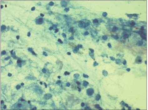
Bronchoalveolar lavage performed the next day showed diffuse blood in the tracheobronchial tree and numerous hemosiderin-laden macrophages (B). Results of testing for glomerular basement membrane (GBM) antibody were normal. A test for c-antineutrophil cytoplasmic antibodies (c-ANCA) was positive; however, the lack of other clinical signs/symptoms suggested that this may have been a nonspecific marker for an autoimmune process; the lung biopsy confirmed the result. The patient received a blood transfusion and remained stable during her 4-day hospital stay. At discharge, her respiratory symptoms had resolved and her hemoglobin level had normalized.
PH can be primary or secondary to an underlying systemic disease. It can be classified into 3 groups: PH associated with circulating anti-GBM antibodies, as seen in Goodpasture syndrome (group 1); PH in immune complex disease, as seen in systemic lupus erythematosus, Henoch-Schönlein purpura, and Heiner syndrome (group 2); and idiopathic PH (group 3).1
Heiner syndrome occurs in children aged 6 months to 2 years. Presenting symptoms may include hemoptysis, iron deficiency anemia, and pulmonary infiltrates. Patients may have prolonged exposure to cow’s milk and a history of chronic rhinitis, recurrent otitis media, persistent cough, or failure to thrive.
The immune-mediated response in Heiner syndrome is not completely understood. The reaction may be secondary to immune complex formation, as in a type 3 hypersensitivity reaction, or it may be cell-mediated. Affected patients also have high levels of circulating IgG against bovine milk.2 It has been postulated that milk aspiration serves as a trigger for the immune-related reaction.3
A complete blood cell count and IgE levels may be beneficial in diagnosing Heiner syndrome. PH in early infancy is associated with eosinophilia, elevated IgE levels, chronic nasal discharge, diarrhea, and upper airway obstruction.4 Tests for anti-GBM and c-ANCA antibodies are helpful in ruling out other causes of PH.5 Chest radiography may be followed by a CT scan if a secondary bacterial infection is suspected. Diagnosis is confirmed by the presence of hemosiderin-laden macrophages on bronchoalveolar lavage.
Treatment consists primarily of strict avoidance of the offending food, in this case cow’s milk. Substitute formulas may be given, such as soy formula. In acute episodes, supplemental oxygen or mechanical ventilation may be required. Corticosteroids remain the first line of treatment. In more severe cases, immunosuppressive therapy with agents such as cyclophosphamide( may be indicated. Without treatment, PH can progress to pulmonary fibrosis and ultimately respiratory failure.
Pulmonary disease caused by milk allergy may be a rare presentation but should be thought of in a child with recurrent infections and pulmonary infiltrates. Patients with PH as a result of exposure to cow’s milk have a better prognosis than do those with other forms of hemosiderosis.


