Hyphema in Spondyloepiphyseal Dysplasia Congenita
Photoclinic
Foresee Your Next Patient
An 8-year-old boy presented to our emergency department with a chief complaint of sudden-onset pain and blindness in his right eye. He had had no symptoms the previous night but had awakened with the sudden pain and blindness.

There was no eye discharge, fever, trauma, or foreign body sensation in the eye. He had no history of prolonged bleeding or excessive bruising. His past medical history was significant for bilateral hip dysplasia and myopia, for which he wears glasses. Family history was significant for a sister who had been diagnosed with arthritis at 12 years of age and a brother who had bilateral hip replacement at 10 years of age.
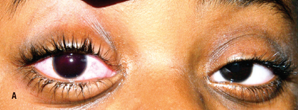
On physical examination, the boy’s vital signs were normal, his height was at the 25th percentile, and his weight was at the 75th percentile. Examination showed a red right eye with a complete hyphema obscuring the pupil (A). Pupillary size and reaction were difficult to assess in the right eye. The conjunctiva was injected, and the patient was unable to see light or hand movements in the affected eye. No conjunctival hemorrhage or discharge was noted, and there was neither proptosis nor pain with eye movements. The left eye was entirely normal on examination, and the rest of his physical examination was unremarkable.
Results of laboratory evaluation, including complete blood count, basic metabolic profile, and coagulation studies, were within normal limits. An ultrasonogram of the right eye done by a pediatric radiologist showed blood in the anterior and posterior chambers, with lens displacement and retinal detachment (B).
The combination of hip dysplasia, retinal detachment, myopia, and a history of siblings with hip and joint disease suggested the diagnosis of spondyloepiphyseal dysplasia congenita, a rare autosomal dominant disorder caused by multiple mutations to COL2A1 gene, which affects collagen-containing tissues. The 2 types of the disease are the mild coxa vara form, with height maintained (as in our patient) and the severe coxa vara form, with clinical short stature. Patients with the severe form usually present at age 3 to 4 years with skeletal deformities and/or hip or spinal dysplasia.

Figure – Anteroposterior right-eye ultrasonogram showing blood in the anterior and posterior chambers, along with lens dislocation (A) and retinal detachment (B).
The affected area determines treatment modality: Bracing is recommended for scoliosis of less than 40 degrees; spinal fusion may be warranted for cases with atlantoaxial instability or severe scoliosis. Children with spondyloepiphyseal dysplasia congenita also may experience hypotonia, progressive weakness, and respiratory insufficiency. Regular pediatric neurology and pulmonology follow-up is recommended. The eye manifestations include myopia and retinal detachment, the latter of which has been reported in up to 50% of patients with the condition. Hearing loss has been reported in 25% of patients. The level of severity of these manifestations varies.
Proper counseling is warranted, along with genetic, ophthalmologic, and orthopedic evaluation. Persons with spondyloepiphyseal dysplasia congenita usually have normal lifespan, albeit with increased morbidity. Our patient had bilateral eye surgery to prevent further retinal detachment, with aspiration of the blood from the right eye.


