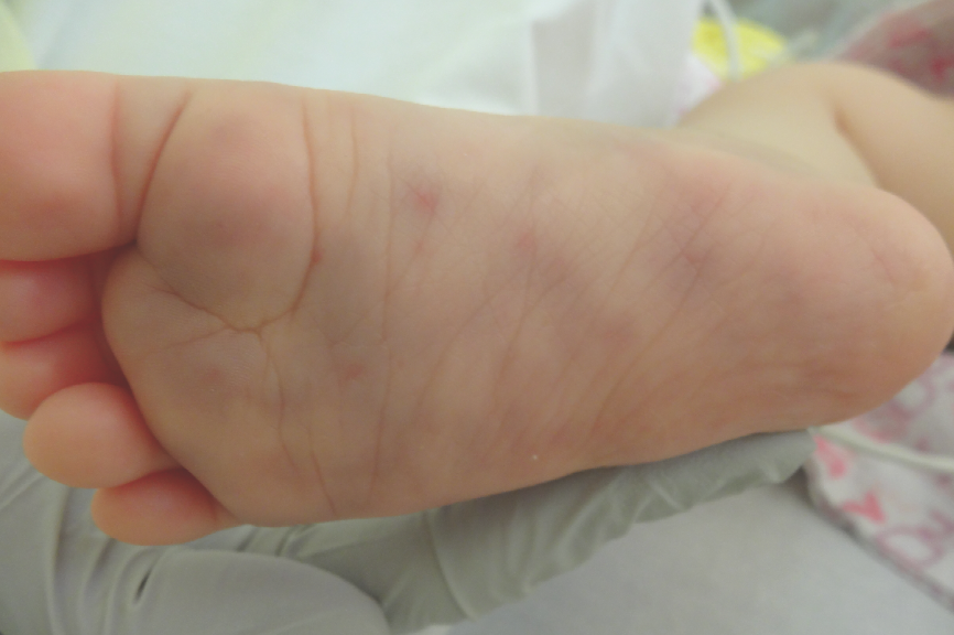
A Suspected Case of Meningococcemia in a 7-Month-Old Girl
A 7-month-old girl presented to the emergency department in the early afternoon with an abrupt onset of excessive sleepiness, new fever, and a rash on the lower extremities.
The mother said that the infant had been “clingy” the day before and had slept through the night, which was unusual for her, but she added that the girl had seemed well that morning before being dropped off at daycare. While at daycare, the infant had become so sleepy that her mother had been called to pick her up. She was taken to her primary care physician, where she was noted to have a fever of 38.8°C. Purplish spots developed over her calves and the dorsa and soles of her feet while she was being examined.
 In the emergency department, physical examination revealed a stable but ill-appearing infant with petechiae and nonpalpable purpura ranging from 6 to 12 mm scattered over both lower extremities, most prominently over the extensor surfaces of her lower extremities and the soles of the feet. New petechiae also were noted along the infant’s neck and cheek and over the labia. There was no conjunctivitis, pallor, mucous membrane change, lymphadenopathy, organomegaly, mottling, or active bleeding from any site. Physical examination findings were otherwise unremarkable.
The girl had had no recent illnesses or sick contacts. Her immunizations were up to date, and she had a normal birth and newborn history.
Laboratory evaluation results demonstrated leukocytosis, with a white blood cell count of 20,600/µL (56% granulocytes). Results of a hemoglobin test, a platelet count, coagulation studies, urinalysis, and cerebrospinal fluid (CSF) analysis were unremarkable. Blood and CSF fluid cultures that were obtained later, after antibiotics were administered, remained sterile.
After consultation with experts in infectious disease and dermatology, the infant received a diagnosis of suspected meningococcemia. She received an intramuscular dose of ceftriaxone empirically, followed by definitive therapy with cefotaxime for 5 to 7 days. Her clinical condition improved rapidly, and she was discharged home without complications.
Her daycare contacts were treated with rifampin; since she was still breastfeeding, the mother was treated with intramuscular ceftriaxone rather than the ciprofloxacin that usually is recommended for adults. The infant’s medical contacts did not require prophylaxis, since they had not been in close contact with her oral secretions.
 Discussion
Meningococcal disease affects approximately 1,000 people per year in the United States, with peak incidence occurring in infants younger than 1 year of age.1 Invasive meningococcal disease in the United States most commonly is caused by serotype B.1
In one study, parents of children with meningococcal disease could pinpoint the precise time at which their child became ill, with progression to unconsciousness, seizures, or death occurring within 24 hours.2 The classic findings of nonblanching rash and meningism are relatively late findings compared with the presence of early signs of sepsis (cool extremities, skin mottling, and leg pain), which occur at a median of 7 to 12 hours.2
 The nonblanching rash typical of meningococcemia is most evident on the trunk and lower extremities and is present in up to 70% of cases. Petechiae can be present in a variety of disease processes; however, meningococcal disease should be suspected when petechiae are seen in the presence of an ill-appearing child, purpura (>2 mm), or delayed capillary refill time (positive likelihood ratios 4.2, 6.9, and 5.5, respectively).3 Meningococcemia is less likely when petechiae are seen in a well-appearing child without purpura, especially if the petechiae are limited to the superior vena cava distribution.3
Diagnosis of invasive Neisseria meningitidis is made by culture of the Gram-negative diplococci in blood, CSF, skin biopsy, or synovial fluid cultures in clinically compatible cases.4 Blood cultures are positive in 50% to 60% of cases but are rapidly sterilized by antibiotics.5 Real-time polymerase chain reaction testing shows promise for detecting disease in patients who have been pretreated with antibiotics, with a sensitivity of 96% compared with blood and CSF cultures.5 Latex agglutination testing has limited usefulness in this age group because of its low sensitivity for detecting serogroup B meningococci.5
Four vaccines are licensed in the United States for prevention of meningococcal disease due to serogroups W, A, C, or Y. Two vaccines for the most prevalent form of disease, caused by serogroup B, are in late-stage clinical development and soon may become available.1
References:
1Cohn AC, MacNeil JR, Clark TA, et al; Centers for Disease Control and Prevention (CDC). Prevention and control of meningococcal disease: recommendations of the Advisory Committee on Immunization Practices (ACIP). MMWR Recomm Rep. 2013;62(RR-2):1-28.
2.Thompson MJ, Ninis N, Perera R, et al. Clinical recognition of meningococcal disease in children and adolescents. Lancet. 2006;367(9508):397-403.
3.Wells LC, Smith JC, Weston VC, Collier J, Rutter N. The child with a non-blanching rash: how likely is meningococcal disease? Arch Dis Child. 2001;85(3):218-222.
4.Meningococcal infections. In: American Academy of Pediatrics. Red Book: 2012 Report of the Committee on Infectious Diseases. 29th ed. Elk Grove Village, IL: American Academy of Pediatrics; 2012:500-509.
5.Apicella M. Diagnosis of meningococcal infection. UpToDate Web site. http://www.uptodate.com/contents/diagnosis-of-meningococcal-infection. Updated January 22, 2014. Accessed February 18, 2014.
Dr Shomaker is a pediatric hospitalist at Children’s Hospital of The King’s Daughters and an assistant professor of pediatrics at Eastern Virginia Medical School in Norfolk, Virginia.


